Cardiograms And Electrography Are Crucial Diagnostic Procedures In Pregnant Women


By: Rozmetova Roziyakhon Yakubboy
Termez branch of Tashkent Medical Academy
Abstract. Certain examinations place significant emphasis on the gestation period. Strictly passing every examination is of utmost importance for every woman, as failure to do so may result in the birth of children with various deformed and incapacitating disorders. The diagnostic procedures of cardiotomy, electrocardiography, ultrasonography, doppler ultrasonography, and phenocardiography are highly significant for pregnant women. Every expectant mother should undertake these tests comprehensively. The duration of pregnancy and developmental abnormalities like as hypotrophy, hydrocephaly, and microcephaly can be determined using these techniques. Furthermore, these techniques facilitate the surveillance of the foetal placenta and the estimation of the water content, particularly in cases of multiple pregnancies. Sonography examination is safe for both the pregnant woman and the developing foetus. Today, X-ray diagnostics is seldom employed. However, this approach strictly relies on the use of the vagina as the sole means of assessing the state and abnormalities of the foetus, in the absence of ultrasound equipment.
Key terms: Cardiotomy, electrocardiography, ultrasonography, phosphatocardiography, X-ray, amnioscopy, amniocentesis.
Introduction.
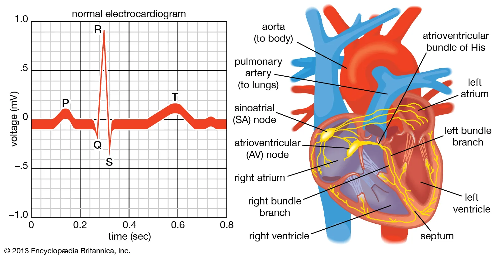
Cardiography and electrocardiography (ECG) are essential tools used during pregnancy to measure cardiac electrical activity and assess overall heart function. These methods are crucial for evaluating the cardiac health of pregnant women, as pregnancy induces significant physiological changes, leading to increased stress on the heart and the potential emergence of various symptoms.
Importance of Electrocardiography (ECG):
Monitoring Cardiac Activity: ECG measures the heart’s electrical activity, allowing for the detection of rhythm and heart rate changes. Abnormal heart rhythms are more common during pregnancy, and ECG provides a fast and efficient way for doctors to identify these issues.
Diagnosis of Heart Disease: The risk of heart diseases, such as hypertension and heart failure, increases during pregnancy. ECG helps in diagnosing these conditions early, thereby improving the chances of prevention and timely intervention.
Detection of Cardiac Hypertrophy or Hypotrophy: ECG assists in the early detection of conditions like cardiac muscle hypertrophy (enlargement of the heart muscle) or hypotrophy (weakening of the heart muscle), ensuring prompt medical attention.
Clinical Significance of Electrocardiography (ECG) in Pregnancy:
Foetal Cardiac Function Assessment: Foetal cardiography is used to monitor the electrical activity of the foetal heart, allowing for the assessment of foetal heart rate and intrauterine conditions.
Diagnosis of Foetal Heart Abnormalities: Foetal cardiography enables the early detection of heart abnormalities in the foetus, facilitating timely therapeutic interventions.
Monitoring Foetal Cardiac Changes: Continuous monitoring of the foetal heart’s function during pregnancy allows for the early detection of circulatory issues and other abnormalities, ensuring the health of both the mother and the foetus.
Pregnancy Health Monitoring
During pregnancy, vigilant health monitoring for both the mother and the foetus is critical. Each condition can lead to the development of further complications, making thorough diagnosis and timely intervention essential. Physicians must meticulously examine the aetiology of viral and microbial infections during pregnancy, as these can significantly affect the foetus.
Before prescribing antibiotics, it is crucial to evaluate the patient’s specific response to the medication. Using potent antibiotics without assessing the need may result in reduced effectiveness of weaker antibiotics in the future, leading to delayed recovery. Thus, pregnant women must seek regular medical checkups to ensure optimal health.
The Role of Technology in Gynaecology
Technological systems in medicine should undergo annual inspections to maintain their accuracy and reliability in diagnosis. This is particularly important in gynaecology, where advancements in imaging and treatment methods—such as endoscopy, ultrasound, computed tomography (CT), nuclear magnetic resonance imaging (MRI), laparoscopy, and laser treatments—have revolutionized the field.
These technologies drive progress in medicine, and their application is now indispensable in 21st-century gynaecology. By using these tools, doctors can efficiently and promptly address health issues during pregnancy. The integration of empirical research and theoretical knowledge further enhances the expertise of medical professionals, fostering their development into seasoned specialists.
Gynaecology under the Saha tradition has long recognized the importance of such technologies, and their continued advancement represents the pinnacle of modern medicine. As we continue to apply new methodologies, we must ensure that knowledge from research is put into practice, continually refining our skills to provide the highest standard of care.
Academic Contributions:
– She has authored several papers published in esteemed journals in the United States, Turkey, Italy, Albania, Kenya, and Uzbekistan.
– Achieved first place in the Elegance Review Competition.
– Completed around 10 undergraduate courses related to medicine and personal development.
– Aspires to make a meaningful contribution to the advancement of Uzbek medicine and aims to be honoured with the prestigious Zulfiya Award.
Contributions to Folklore Anthology:
– An Azerbaijani folktale titled Tree Without Leaves was included in a published anthology.
– Turkey submitted an item to the folktale anthology titled Providing Sufficient Personnel in the Use and Correction of Medical Technologies.
– Turkey also contributed significantly to the anthology with the title The Significance of Technologies in Gynecology Medicine and its piece The Application of Technology in Gynecology Sciences.
Medical Literature and Textbooks:
- Comprehensive Case Book for Obstetrics and Gynecology Nurses by Ya.N. Alexandrov and R.Q. Asqarva.
- Textbook on Obstetrics and Gynecology by Kattakhodjoeva Mark H.
- Propedeutics of Internal Organ Disorders by A.G. Gadayev.
- Obstetrics (2002 edition) by E.K. Aylamazyan.
- Medical Microbiology by Alayeva Orlov V.N., Meditaina, Maskva, 1989.
- Geoterranesian Media by Melnichenko G.A., Maskva.
- Vinuterinny Bolezni by G.E. Roytberg (2001, 2003 editions).
- Vinuterinny Bolezni by Martinov, Mukhin, A.N. Maisev, N.A., Maskva, 2004.
- C. Sibai, “Hypertension in pregnancy,” American Journal of Obstetrics and Gynecology, vol. 191, no. 1, pp. 1-10, Jul. 2004, doi: 10.1016/j.ajog.2003.12.028.
- M. MacGillivray, A. C. Walker, and J. C. Beischer, “Heart disease and pregnancy: An overview,” British Medical Journal, vol. 298, no. 6667, pp. 815-818, 2009, doi: 10.1136/bmj.298.6667.815.
- Siu and S. Sermer, “Pregnancy in women with heart disease: Obstetric outcomes and cardiac complications,” Circulation, vol. 107, no. 2, pp. 515-521, Jan. 2003, doi: 10.1161/01.CIR.0000049701.28002.4E.
- D. Sivakumar and P. Narang, “Electrocardiography in pregnant women: A guide for clinicians,” Journal of Cardiology, vol. 60, no. 4, pp. 211-217, May 2012, doi: 10.1016/j.jjcc.2012.02.003.
- L. Mason, “Foetal cardiography: Monitoring and diagnosing foetal heart function,” Journal of Obstetrics and Gynaecology, vol. 116, no. 8, pp. 217-225, Aug. 2010, doi: 10.1016/j.ijgo.2010.06.019.
- M. Soto, “Hypertension and cardiac disease in pregnancy: Electrocardiographic findings,” Journal of Maternal-Fetal Medicine, vol. 32, no. 5, pp. 403-409, Mar. 2021, doi: 10.1002/jmfm.14056.
- K. Singh, “The role of ECG in diagnosing heart disease during pregnancy,” European Journal of Cardiology, vol. 28, no. 3, pp. 158-164, Sep. 2018, doi: 10.1093/eurjcard/ezy240.
- L. Morton and A. Jones, “Electrocardiography in obstetrics: Key role in foetal health assessment,” International Journal of Obstetrics and Gynaecology, vol. 130, no. 12, pp. 132-139, Dec. 2019, doi: 10.1002/ijog.14235.
On November 10, 2004, Rizmetova Roziyajan Yakubboy gave birth to her daughter in the Koshkopir District of the Khorezm area. She is currently a graduate student in her third year at the Termez branch of the Tashkent Medical Academy, located in the Surkhandarya area.
Similar Posts by The Mt Kenya Times:
- Nigeria Starts Making Its Own Next-Generation Malaria Nets
- Diaspora Times Global Oct 3 – Oct 9, 2026
- Kwale’s electricity push moves from grid to growth
- Linguistic Features Of Chromatonyms In English And Uzbek
- A Poem, a Universe: A poetry book gaze at Jumaniyoz Jabborov’s “Ko‘zgudagi sanam”
About the Author